What to know about cardiac muscle tissue
Cardiac muscle tissue, or myocardium, is a specialized type of muscle tissue that forms the heart. This muscle tissue, which contracts and releases involuntarily, is responsible for keeping the heart pumping blood around the body. The human body contains three different kinds of muscle tissue: skeletal, smooth, and cardiac. Only cardiac muscle tissue, comprising cells called myocytes, is present in the heart. In this article, we discuss the structure and function of cardiac muscle tissue. We also cover medical conditions that can affect cardiac muscle tissue and tips for keeping it healthy.
A person can strengthen cardiac muscle tissue by doing regular exercise.
Muscle is fibrous tissue that contracts to produce movement. There are three types of muscle tissue in the body: skeletal, smooth, and cardiac. Cardiac muscle is highly organized and contains many types of cell, including fibroblasts, smooth muscle cells, and cardiomyocytes.

Cardiac muscle only exists in the heart. It contains cardiac muscle cells, which perform highly coordinated actions that keep the heart pumping and blood circulating throughout the body. Unlike skeletal muscle tissue, such as that which is present in the arms and legs, the movements that cardiac muscle tissue produces are involuntary. This means that they are automatic, and that a person cannot control them.
The heart also contains
specialized types of cardiac tissue containing "pacemaker" cells.
These contract and expand in response to electrical impulses from the nervous
system. Pacemaker cells generate electrical impulses, or action potentials,
that tell cardiac muscle cells to contract and relax. The pacemaker cells
control heart rate and determine how fast the heart pumps blood.
How is it structured?
Cardiac muscle tissue gets its strength and flexibility from its interconnected cardiac muscle cells, or fibers.
Most cardiac muscle cells contain one nucleus, but some have two. The nucleus houses all of the cell's genetic material.
Cardiac muscle cells also contain mitochondria, which many people call "the powerhouses of the cells." These are organelles that convert oxygen and glucose into energy in the form of adenosine triphosphate (ATP).
Cardiac muscle cells appear striated or striped under a microscope. These stripes occur due to alternating filaments that comprise myosin and actin proteins. The dark stripes indicate thick filaments that comprise myosin proteins. The thin, lighter filaments contain actin.
When a cardiac muscle cell contracts, the myosin filament pulls the actin filaments toward each other, which causes the cell to shrink. The cell uses ATP to power this contraction.
A single myosin filament connects to two actin filaments on either side. This forms a single unit of muscle tissue, called a sarcomere.
Intercalated discs connect cardiac muscle cells. Gap junctions inside the intercalated discs relay electrical impulses from one cardiac muscle cell to another.
Desmosomes are other structures present within intercalated discs. These help hold cardiac muscle fibers together.


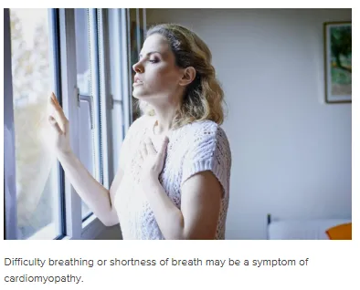
Difficulty breathing or shortness of breath may be a symptom of cardiomyopathy.
Cardiomyopathy refers to a group of medical conditions that affect cardiac muscle tissue and impair the heart's ability to pump blood or relax normally.
Some common symptoms of cardiomyopathy include:
· difficulty breathing or shortness of breath
· fatigue
· swelling of the legs, ankles, and feet
· inflammation in the abdomen or neck
· irregular heartbeat
· heart murmurs
· dizziness or lightheadedness
Factors that can increase a person's risk of cardiomyopathy include:
· diabetes
· thyroid disease
· coronary heart disease
· heart attack
· chronic high blood pressure
· viral infections that affect the heart muscle
· valvular disease of the heart
· heavy alcohol consumption
· a family history of cardiomyopathy
A heart attack due to a blocked artery can cut off the blood supply to certain areas of the heart. Eventually, the cardiac muscle tissue in these areas will start to die. The death of cardiac muscle tissue can also occur when the heart's oxygen demand exceeds the oxygen supply. This causes the release of cardiac proteins such as troponin into the bloodstream.
Some examples of cardiomyopathy include:
Dilated cardiomyopathy
Dilated cardiomyopathy causes the cardiac muscle tissue of the left ventricle to stretch and the heart's chambers to dilate.
Hypertrophic cardiomyopathy
Hypertrophic cardiomyopathy (HCM) is a genetic condition in which the cardiomyocytes are not arranged in a coordinated fashion and are instead disorganized. HCM can interrupt blood flow out of the ventricles, cause arrhythmias (abnormal electrical rhythms), or lead to congestive heart failure.
Restrictive cardiomyopathy
Restrictive cardiomyopathy (RCM) refers to when the walls of the ventricles become stiff. When this happens, the ventricles cannot relax enough to fill with an adequate amount of blood.
Arrhythmogenic right ventricular dysplasia
This rare form of cardiomyopathy causes fatty infiltration in cardiac muscle tissue in the right ventricle.
Transthyretin amyloid cardiomyopathy
Transthyretin amyloid cardiomyopathy (ATTR-CM) develops when amyloid proteins collect and form deposits in the walls of the left ventricle. The amyloid deposits cause the ventricle's walls to stiffen, which prevents the ventricle from filling with blood and reduces its ability to pump blood out of the heart. This is a form of RCM.

Children should do 60 minutes of moderate- to high-intensity physical activity each day.
Doing regular aerobic exercise can help strengthen the cardiac muscle tissue and keep the heart and lungs healthy.
Aerobic activities involve moving the large skeletal muscles, which causes a person to breathe faster and their heartbeat to quicken.
Doing these types of activities often can train the heart to become more efficient.
Some examples of aerobic exercises include:
· running or jogging
· walking or hiking
· cycling
· swimming
· jumping rope
· dancing
· jumping jacks
· climbing stairs
The Department of Health and Human Services (DHHS) make the following recommendations in their Physical Activity Guidelines for Americans:
· Children aged 6–17 years old should do 60 minutes of moderate- to high-intensity physical activity each day.
· Adults aged 18 years and older should do 150 minutes of moderate-intensity, or 75 minutes of high-intensity, aerobic exercise each week.
· Pregnant women should try to do at least 150 minutes of moderate-intensity aerobic activity per week.
The DHHS also suggest that a person should try to spread aerobic activity throughout the week. Adults with chronic conditions or disabilities can replace aerobic exercise with at least two muscle-strengthening sessions per week.