PRINCIPLES OF SOUND
Sound is a form of mechanical energy that travels in a longitudinal wave in a series of compressions (high pressure) and rarefactions (low pressure). The transmission of sound requires a medium such as air, liquid, or tissue. Because all mediums have some elastic properties, energy and momentum are transported by means of disturbance from one point to another, without the transport of matter.
Wavelength and frequency are terms often used to describe the relationship between the compressions and rarefactions. The area that includes one complete compression and rarefaction is called a cycle. Wavelength (λ) is the distance over which the acoustic disturbance repeats itself at any instant of time during the cycle. It is summarized by the equation λ = c/f, where f is the frequency and c is the speed of sound. The period of a wave is the time it takes for one cycle to occur. Frequency is the number of cycles that occur per second, and is measured in Hertz (Hz).
Human hearing has a limited range of 20 Hz to 20 kilohertz (kHz). Ultrasound is considered those frequencies greater than 20 kHz, while diagnostic ultrasound utilizes frequencies greater than 1 megahertz (MHz). Amplitude (A) is the maximum increase (or decrease) in pressure due to the presence sound. It is often described as the “height” of the wave, and has no relationship to the wavelength or frequency.
Anatomy of a sound wave.
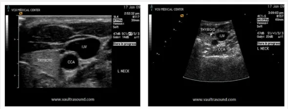
Frequency is important factor in ultrasound imaging. High frequency transducers (> 7MHz) have better resolution at shallow depths, but have limited imaging capability the deeper the wave penetrates the tissue. Lower frequencies allow for better imaging of deeper structures, but with poorer image resolution. Both images below are of the left neck. The image on the left was generated by a 9 MHz transducer creating a shallow (approximately 3cm in depth), but high resolution image of the internal jugular vein (IJV), common carotid artery (CCA) and thyroid gland. The image on the right shows the same structures imaged with a lower frequency (4MHz) transducer. Although the image depth has increased to 6cm, the resolution is greatly diminished.

Comparison of high (L) and low (R) frequency ultrasound imaging of the left neck.
Sound travels at different speeds through different medium. This is known as propagation velocity. In diagnostic ultrasound, human tissue is the medium in which sound travels. The standardized propagation velocity of sound through tissue is approximately 1,540 m/s,which is derived by averaging the different velocities of soft tissues in the body. By comparison, the propagation velocity of air is quite slow at 331 m/s and in bone it can be as fast as 5000 m/s. Table One lists propagation velocities for various tissues in the body.
Propagation velocities of different mediums.