Connective Tissues
Connective tissues are made up of a matrix consisting of living cells and a non-living substance, called the ground substance. The ground substance is made of an organic substance (usually a protein) and an inorganic substance (usually a mineral or water). The principal cell of connective tissues is the fibroblast. This cell makes the fibers found in nearly all of the connective tissues. Fibroblasts are motile, able to carry out mitosis, and can synthesize whichever connective tissue is needed. Macrophages, lymphocytes, and, occasionally, leukocytes can be found in some of the tissues. Some tissues have specialized cells that are not found in the others. The matrix in connective tissues gives the tissue its density. When a connective tissue has a high concentration of cells or fibers, it has proportionally a less dense matrix.
The organic portion or protein fibers found in connective tissues are either collagen, elastic, or reticular fibers. Collagen fibers provide strength to the tissue, preventing it from being torn or separated from the surrounding tissues. Elastic fibers are made of the protein elastin; this fiber can stretch to one and one half of its length and return to its original size and shape. Elastic fibers provide flexibility to the tissues. Reticular fibers are the third type of protein fiber found in connective tissues. This fiber consists of thin strands of collagen that form a network of fibers to support the tissue and other organs to which it is connected. The various types of connective tissues, the types of cells and fibers they are made of, and sample locations of the tissues is summarized in Table 14.3.
|
Table 14.3. |
|||
|
Connective Tissues |
|||
|
Tissue |
Cells |
Fibers |
Location |
|
loose/areolar |
fibroblasts, macrophages, some lymphocytes, some neutrophils |
few: collagen, elastic, reticular |
around blood vessels; anchors epithelia |
|
dense, fibrous connective tissue |
fibroblasts, macrophages, |
mostly collagen |
irregular: skin regular: tendons, ligaments |
|
cartilage |
chondrocytes, chondroblasts |
hyaline: few collagen fibrocartilage: large amount of collagen |
shark skeleton, fetal bones, human ears, intervertebral discs |
|
bone |
osteoblasts, osteocytes, osteoclasts |
some: collagen, elastic |
vertebrate skeletons |
|
adipose |
adipocytes |
few |
adipose (fat) |
|
blood |
red blood cells, white blood cells |
none |
blood |
Loose/Areolar Connective Tissue
Loose connective tissue, also called areolar connective tissue, has a sampling of all of the components of a connective tissue. As illustrated in Figure 14.12, loose connective tissue has some fibroblasts; macrophages are present as well. Collagen fibers are relatively wide and stain a light pink, while elastic fibers are thin and stain dark blue to black. The space between the formed elements of the tissue is filled with the matrix. The material in the connective tissue gives it a loose consistency similar to a cotton ball that has been pulled apart. Loose connective tissue is found around every blood vessel and helps to keep the vessel in place. The tissue is also found around and between most body organs. In summary, areolar tissue is tough, yet flexible, and comprises membranes.

Figure 14.12. Loose connective tissue is composed of loosely woven collagen and elastic fibers. The fibers and other components of the connective tissue matrix are secreted by fibroblasts.
Fibrous Connective Tissue
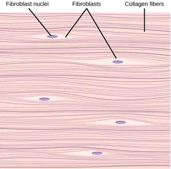
Fibrous connective tissues contain large amounts of collagen fibers and few cells or matrix material. The fibers can be arranged irregularly or regularly with the strands lined up in parallel. Irregularly arranged fibrous connective tissues are found in areas of the body where stress occurs from all directions, such as the dermis of the skin. Regular fibrous connective tissue, shown in Figure 14.13, is found in tendons (which connect muscles to bones) and ligaments (which connect bones to bones).

Figure 14.13. Fibrous connective tissue from the tendon has strands of collagen fibers lined up in parallel.