Exploring Brain Function with Magnetic Resonance Imaging
Advancements in brain imaging using non-invasive technologies such as functional magnetic resonance imaging (fMRI), magnetoencephalography (MEG) and electroencephalography (EEG) have allowed neuroscientists to obtain a greater understanding on how the brain functions with its environment.
Understanding neuro-networks will assist in the development of treatments towards neurodegenerative diseases such as Alzheimer’s disease and Parkinson’s disease. In order to complement brain imaging, various animal models have been developed to investigate the genetic nature of disease states, especially concentrating on neurophysiological processes.
A useful brain imaging technique uses functional magnetic resonance imaging (fMRI) by analysing metabolic changes such as blood oxygenation. The advantage of using fMRI is that it produces good spatial resolution. The disadvantages of fMRI is in its poor time resolution of approximately six seconds and is therefore too slow for tracking single or clusters of neurons in real time. This is because the change in blood flow takes several seconds to catch up with the neural activity. However, fMRI has good spatial specificity in the areas of localised brain function compared to EEG which uses electrical signals and the magnetic signals derived from MEG.
Consequently, both EEG and MEG have an excellent temporal resolution but very poor spatial precision. This is even though these imaging techniques can track neuron population activity within several hundreds of milliseconds. During these processes, both imaging modalities cannot distinguish which set of neurons are being used.
In order to circumvent these issues, fMRI is sometimes used in conjunction with EEG to obtain paramount temporal and spatial precision.
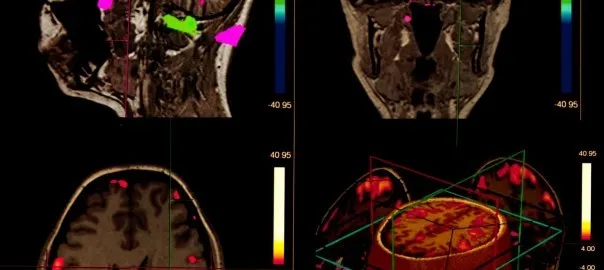
Brain imaging can make a contribution to the patient through personalised medicine in the treatment and management of neurological diseases by creating 3-D individual images in real time. Both MRI and computed tomography (CT) hybrid scanners can be used to generate 3-D images of the brain at a specific moment.
These 3-D images of brain volume are made up of voxels.
The spatial resolution of the MRI scanner determines how the small voxels can be measured during brain imaging of the neural networks. Therefore, stronger magnetic field strength will increase the spatial resolution and will enable better resolution of brain structure.
Image-guided systems controlled by advanced brain navigation software will assist neurosurgeons in precise locations to perform the operation on the patient. These systems are based on the Talairach coordinates (x, y and z).
Comparison of Neuroimaging Modalities
IMAGING MODALITY | RESOLUTION | APPLICATION | ADVANTAGES | DISADVANTAGES |
FUNCTIONAL IMAGING METHODS
| ||||
EEG | S – LOW T – HIGH | Study various rhythms, epilepsy, preoperative mapping, degenerative disorders | Non-invasive, no ionising radiation, widely used, low cost | Low spatial resolution |
MEG | S – MEDIUM T – HIGH | Study epilepsy | Non-invasive, no ionising radiation, can identify epileptic foci | Low spatial resolution |
fMRI | S – LOW T – HIGH | Preoperative mapping, functional mapping | Non-invasive, no ionising radiation | High cost |
S = Spatial Resolution; T = Temporal Resolution