 Figure 1: Picture of a CT Scanner
What is medical imaging?
Medical imaging refers to techniques and processes used to create images of various parts of the human body for diagnostic and treatment purposes within digital health.
The term, medical imaging, includes various radiological imaging techniques such as:
· X-ray radiography
· Fluoroscopy
· Magnetic resonance imaging (MRI)
· Medical ultrasonography or ultrasound
· Endoscopy
· Elastography
· Tactile imaging
· Thermography
· Medical photography and nuclear medicine functional imaging techniques e.g. positron emission tomography (PET)
Medical Imaging is the use of imaging modalities and processes to get pictures of the human body, which can assist diagnosis and treatment of patients. It can also be used to track any ongoing issues, and can therefore help with treatment plans.
There are many different types of medical imaging techniques, which use different technologies to produce images for different purposes. This page will give an introduction to the most common imaging techniques, and the page on uses of AI in radiology will show how some of these techniques, combined with AI, will pave the way for more accurate imaging.
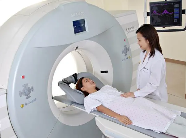
A computerised tomography scan, more commonly called a CT scan, can create a detailed image of the inside of the body using x-rays and computers. It is different to an x-ray because it produced a cross-sectional image of the body, similar to an MRI, making them better at looking at soft tissue and more subtle parts of the image that an x-ray may not pick up.
They can be used to visualise bones, internal organs and blood vessels. Upper body, such as the brain, neck, spine, chest and sinuses are commonly scanned.
They are frequently used in diagnosis, for example to find tumours, or to see broken bones. Another use is to find more detail after another scan, such as an x-ray. Monitoring is also an important use of CT scanners, as regular scans allow progress to be kept of any developing conditions, e.g. cancer.
As shown in the image below, the patient lies on their back on a panel. This panel passes into the scanner, which rotates around the section of your body currently in the scanner. The patient needs to lay still so that the scan can get a clear image. Usually, the radiologist who is operating the machine stands in another room to avoid the radiation, but can communicate with the patient via an intercom. The scan can take from 10 to 20 minutes, but results are available as soon as a computer has analysed the scans.
Figure 1: Picture of a CT Scanner
· CT scans are fairly short - they only last about 10 - 20 minutes.
· The results are extremely fast compared to some other types of scans.
· CT scans are painless, as they are non-invasive.
· As with lots of scans, your body is exposed to some radiation. The more of the patient’s body that is scanned, the more radiation they are exposed to. However, they are designed to minimise radiation exposure.
· There is the possibility of an allergic reaction to the dye used.
A magnetic resonance imaging scan, more commonly known as an MRI scan, is a detailed cross-sectional image of a part of the body. It is similar to a CT scan, but has a higher quality, so it is easier to see differences in tissues, as shown in the picture below.
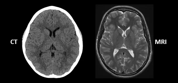 Figure 2: A CT scan vs an MRI scan
MRIs can be used to get images of the brain and spinal cord, bones, the heart, blood vessels and different internal organs.
The uses are similar to those of a CT scanner: diagnosis, getting more detail for treatment planning, and monitoring of ongoing treatment.
Unlike a CT scanner, an MRI surrounds the whole body. The patient is pushed into a thin tube, about 24 inches in diameter, and extremely strong magnets and radio waves are used to created detailed images. Similarly again to a CT scanner, the radiographer will stand in another room viewing the results and communicating via an intercom, but the MRI is much louder than a CT scanner. They can take from 15 to 90 minutes.
· MRI scans are painless and safe, as the magnetic fields and radio waves have no known negative impact on the patient.
· They don’t involve any x-ray radiation exposure, so can be used by pregnant women and babies if necessary.
· MRI scans enclose lots of the body, so make people with claustrophobia uncomfortable.
· Metal cannot go inside of an MRI scanner, so people with certain implants such as pacemakers cannot use them.
A PET scan can create a 3D image of the inside of the body. They can be combined with CT and MRI scans to create an even clearer image to show what is going on. They can also be focused on specific parts of the body, and show how well a part of the body is working. The image below shows how a PET scan and CT scan can be combined.
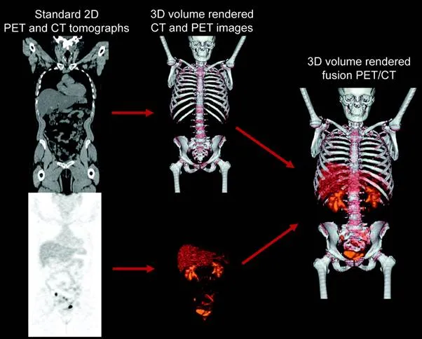 Figure 3: Combination of a CT scan and a PET scan
They are used to detect the progress of cancer, and can be used to get high resolution images of the brain.
They are commonly used in people who have already been diagnosed with cancer, as they can clearly show how far a cancer has spread or responded to treatments such as chemotherapy. They are also used in the planning of surgery, such as brain or heart operations. Dementia can also be diagnosed with a PET scan, as it can show if the brain’s normal function is changed.
A radiotracer, usually fluorodeoxyglucose (FDG), is injected into your arm, and this gives off radiation. The PET scanner can detect this radiation when it collects in certain parts of your body. If there is an area where the FDG is not building up, then there is a certain body function which isn’t working there. Cancerous cells use glucose at a faster rate than normal, and therefore by checking the FDG concentration, cancer can be identified and tracked in the body. The PET scanner machine looks similar to an MRI machine. The scan takes about 30 minutes.
· The radiotracer fluorodeoxyglucose is used is similar to glucose, so the body treats it in a similar way.
· The scan only takes about 30 minutes.
· The PET scan can reveal cell level metabolic changes occurring in an organ or tissue, which a CT or MRI cannot.
· The PET scan exposes you to radiation, which may lead to cancer. However, the amount is quite small. The radioactive tracer has a short half life.
· Patients should avoid people who shouldn’t be exposed to radiation, such as pregnant women, for a few hours after the scan.
An ultrasound uses high-frequency waves to show what is inside a part of the body. It is also known as a sonogram.
Ultrasounds can produce images of unborn children in real time, as shown below.
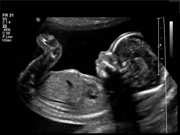 Figure 4: An ultrasound image of an unborn baby
The most common use are to monitor unborn babies, however they are also used in diagnosis and during certain procedures for guiding surgeons.
The device has a probe, which emits high-frequency sound waves. They bounce off of different parts of the body, creating echoes, and when these bounce back to the probe, it can also detect them. This can create a live image on another scanner. The scan can last from 15 to 45 minutes. They can be done externally, internally or endoscopically.
· Usually there are no after-effects of ultrasound scans. This means normal activity can be resumed straight after.
· The results are seen in real time, so there is no need to wait.
· Some probe covers have latex, which can be a problem if the patient is allergic to latex.
· Endoscopic ultrasounds can cause a sore throat or bloating, or in extreme cases, internal bleeding.
An x-ray is a very common procedure used to get images of inside of the body. It uses radiation in the x-ray part of the electromagnetic spectrum.
They are used to produce images of bones, usually to see if and where there are breaks. They are also used by dentists and orthodontists to look at teeth. Bone tumours can also be seen on x-rays.
They can be used to guide surgeons as they are operating. They can also be used to detect broken bones, and to plan the best course of treatment for this.
X-rays, which are a type of electromagnetic radiation invisible to humans, pass through the body. The energy is absorbed by different parts of the body at different rates, and a detector on the other side of the person will see how much was absorbed, and will be able to generate an image from this. Denser parts of the body, such as bone, show up as white, as few x-rays could pass through. Sometimes a contrast agent is given to the patient in order for soft tissues to be seen more easily on the image. The x-ray is extremely fast, and the whole procedure should only take a few minutes.
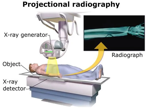 Figure 5: Diagram of how an x-ray scanner works
· The machine does not surround the whole body, so will not cause anxiety in people with claustrophobia.
· The procedure only takes a few minutes.
· Some contrast agents may cause unwanted side effects.
· X-rays expose the patient to unwanted radiation, which could potentially cause cancer, however the amount of radiation given off is minimal.