The Conducting System of the Heart
The cardiac conduction system is a collection of nodes and specialised conduction cells that initiate and co-ordinate contraction of the heart muscle. It consists of:
· Sinoatrial node
· Atrioventricular node
· Atrioventricular bundle (bundle of His)
· Purkinje fibres
Overview of Heart Conduction

Fig 1 – Animation of the spread of conduction through the heart
The sequence of electrical events during one full contraction of the heart muscle:
· An excitation signal (an action potential) is created by the sinoatrial (SA) node.
· The wave of excitation spreads across the atria, causing them to contract.
· Upon reaching the atrioventricular (AV) node, the signal is delayed.
· It is then conducted into the bundle of His, down the interventricular septum.
· The bundle of His and the Purkinje fibres spread the wave impulses along the ventricles, causing them to contract.
We will now discuss the anatomy of the individual components involved in the conducting system.
Components of the Cardiac Conduction System
Sinoatrial Node
The sinoatrial (SA) node is a collection of specialised cells (pacemaker cells), and is located in the upper wall of the right atrium, at the junction where the superior vena cava enters.
These pacemaker cells can spontaneously generate electrical impulses. The wave of excitation created by the SA node spreads via gap junctions across both atria, resulting in atrial contraction (atrial systole) – with blood moving from the atria into the ventricles.
The rate at which the SA node generates impulses is influenced by the autonomic nervous system:
· Sympathetic nervous system – increases firing rate of the SA node, and thus increases heart rate.
· Parasympathetic nervous system – decreases firing rate of the SA node, and thus decreases heart rate.
Atrioventricular Node
After the electrical impulses spread across the atria, they converge at the atrioventricular node – located within the atrioventricular septum, near the opening of the coronary sinus. The AV node acts to delay the impulses by approximately 120ms, to ensure the atria have enough time to fully eject blood into the ventricles before ventricular systole. The wave of excitation then passes from the atrioventricular node into the atrioventricular bundle.
Atrioventricular Bundle
The atrioventricular bundle (bundle of His) is a continuation of the specialised tissue of the AV node, and serves to transmit the electrical impulse from the AV node to the Purkinje fibres of the ventricles.
It descends down the membranous part of the interventricular septum, before dividing into two main bundles:
· Right bundle branch – conducts the impulse to the Purkinje fibres of the right ventricle
· Left bundle branch – conducts the impulse to the Purkinje fibres of the left ventricle.
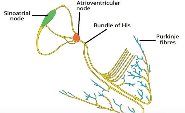
Fig 2 – Overview of the individual components of the heart conduction pathway
Purkinje Fibres
The Purkinje fibres (sub-endocardial plexus of conduction cells) are a network of specialised cells. They are abundant with glycogen and have extensive gap junctions. These cells are located in the subendocardial surface of the ventricular walls, and are able to rapidly transmit cardiac action potentials from the atrioventricular bundle to the myocardium of the ventricles. This rapid conduction allows coordinated ventricular contraction (ventricular systole) and blood is moved from the right and left ventricles to the pulmonary artery and aorta respectively.