Fast Imaging
The 2D FT scan of k-space has the disadvantage that the scan time is proportional to the number of phase encodes. It is often advantageous to trade off SNR for a shorter scan time. This is especially true when motion artifacts dominate thermal noise. To allow for a flexible tradeoff of SNR for imaging time, more than a single line in k-space must be covered in a single excitation. The most popular approach, called echo-planar imaging (EPI), traverses k-space back and forth on a single excitation pulse. The k-space trajectory is drawn in Figure 12.2.
It is important that the tradeoff be flexible so that you can maximize the imaging time given the motion constraints. For example, patients can hold their breath for about 12 sec. So a scan of 12 sec duration gives the best SNR given the breath-hold constraint. The EPI trajectory can be interleaved to take full advantage of the breath-hold interval. If each acquisition takes about a second, 12 interleaves can be collected. Each interleaf acquires every twelfth line in k-space.
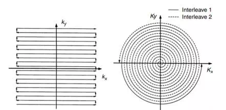
FIGURE 12.2 Alternative methods for the rapid traversal of k-space. On the left is the echo planar trajectory. Data are collected during the horizontal traversals. When all Ny horizontal lines in k-space have been acquired, the data are sent to a 2D FFT to reconstruct the image. On the right is an interleaved spiral trajectory. The data are interpolated to a 2D rectilinear grid and then Fourier transformed to reconstruct the image. These scanning techniques allow for imaging within a breathhold.
Another trajectory that allows for a flexible tradeoff between scan time and SNR is the spiral trajectory. Here the trajectory starts at the origin in k-space and spirals outward. Interleaving is accomplished by rotating the spirals. Figure 12.2 shows two interleaves in a spiral format. Interleaving is very helpful for reducing the hardware requirements (peak amplitude, peak slew rate, average dissipation, etc.) for the gradients amplifiers. For reconstruction, the data are interpolated to a 2D rectilinear grid and then Fourier-transformed. Our group has found spiral imaging to be very useful for imaging coronary arteries within a breath-hold scan . The spiral trajectory is relatively immune to artifacts due to the motion of blood.