Multidetector SPECT System
The first fully functional SPECT imaging acquisition system was designed and constructed by Kuhl and Edwards [Kuhl and Edwards, 1963, 1964, 1968] in the 1960s, well before the conception of x-ray CT. As shown in Figure 13.7a, the MARK IV brain SPECT system consisted of four linear arrays of eight discrete NaI(TI) scintillation detectors assembled in a square arrangement. Projection data were obtained by rotating the square detector array around the patient’s head. Although images from the pioneer MARK IV SPECT system were unimpressive without the use of proper reconstruction methods that were developed in later years, the multidetector design has been the theme of several other SPECT systems that were developed. An example is the Gammatom-1 developed by Cho et al. [1982]. The design concept also was used in a dynamic SPECT system [Stokely et al., 1980] and commercial multidetector SPECT systems marketed by Medimatic, A/S (Tomomatic-32). Recently, the system design was extended to a multislice SPECT system with the Tomomatic-896, consisting of eight layers of 96 scintillation detectors. Also, the system allows both body and brain SPECT imaging by varying the aperture size.
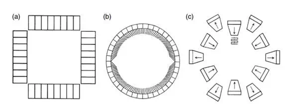
FIGURE 13.7 Examples of multidetector-based SPECT systems. (a) The MARK IV system consists of four arrays of eight individual NaI(TI) detectors arranged in a square configuration. (b) The Headtome-II system consists of a circular ring of detectors. A set of collimator vanes that swings in front of the discrete detector is used to collect projection data from different views. (c) A unique Cleon brain SPECT system consists of 12 detectors that scan both radially and tangentially.
Variations of the multiple-detectors arrangement have been proposed for SPECT system designs. Figure 13.7b shows the Headtome-II system by Shimadzu Corporation [Hirose et al., 1982], which consists of a stationary array of scintillation detectors arranged in a circular ring. Projection data are obtained by a set of collimator vanes that swings in front of the discrete detectors. A unique Cleon brain SPECT system (see Figure 13.7c), originally developed by Union Carbide Corporation in the 1970s, consists of 12 detectors that scan both radially and tangentially [Stoddart and Stoddart, 1979]. Images from the original system were unimpressive due to inadequate sampling, poor axial resolution, and a reconstruction algorithm that did not take full advantage of the unique system design and data acquisition strategy. A much improved version of the system with a new reconstruction method [Moore et al., 1984] is currently marketed by Strichman Corporation.
The advantages of multidetector SPECT systems are their high sensitivity per image slice and high counting rate capability resulting from the array of multidetectors fully surrounding the patient. However, disadvantages of multidetector SPECT systems include their ability to provide only one or a few noncontiguous cross-sectional image slices. Also, these systems are relatively more expensive compared with camera-based SPECT systems described in the next section. With the advance of multicamera SPECT systems, the disadvantages of multidetector SPECT systems outweigh their advantages. As a result, they are less often found in nuclear medicine clinics.