Advances in Functional Brain Mapping
The popularity of fMRI is based on many factors. It is safe and totally noninvasive. It can be acquired in single subjects for a scanning duration of several minutes, and it can be repeated on the same subjects as many times as necessary. The implementation of the blood oxygenation sensitive MR technique is universally available. Early neuroimaging, work focused on time-resolved MR topographic mapping of human primary visual (VI) (Figure 12.15 and Figure 12.16), motor (MI), somatosensory (S1), and auditory (A1) cortices during task activation. Today, with BOLD technique combined with EPI, one can acquire 20 or more contiguous brain slices covering the whole head (3 × 3 mm in plane and 5 mm slice thickness) every 3 sec for a total duration of several minutes. Conventional scanners can only acquire a couple of slices at a time. The benefits of whole-head imaging are many. Not only can researchers identify and test their hypotheses on known brain activation centers, they can also search for previous unknown or unsuspected sites. High resolution work done with EPI has a resolution of 1.5×1.5 mm in plane and a slice thickness of 3 mm. Higher spatial resolution has been reported in conventional 1.5-T MR systems.
Of note with Figure 12.16 is that with blood oxygenation–sensitive MR technique, one observers an undershoot in signal in V1 when the light stimulus is turned off. The physiologic mechanism underlying the undershoot is still not well understood.
The data collected in the last 3 years have demonstrated that fMRI maps of the visual cortex correlate well with known retinotopic organization. Higher visual regions such as V5/MT and motor-cortex organization have been explored successfully. Preoperative planning work (Figure 12.17) using motor stimulation has helped neurosurgeons who attempt to preserve primary areas from tumors to be resected. For higher cognitive functions, several fMRI language studies have already demonstrated known language-associated regions (Figure 12.18). There is more detailed modeling work on the mechanism of functional brain mapping by blood-oxygenation change [43– 46]. Postprocessing techniques that would help to alleviate the serious problem of motion/displacement artifacts are available.
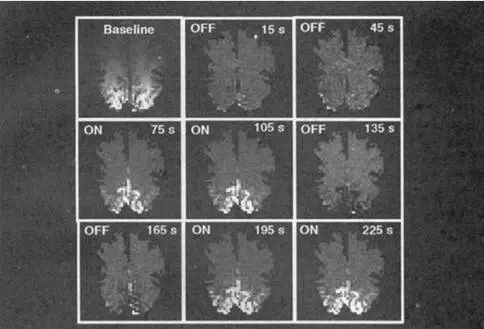
FIGURE 12.15 Movie of fMRI mapping of primary visual cortex (V1) activation during visual stimulation. Images are obliquely aligned along the calcarie fissures with the occipital pole at the bottom. Images were acquired at 3-sec intervals using a blood oxygenation–sensitive MRI sequence (80 images total). A baseline image acquired during darkness (upper left) was subtracted from subsequent images. Eight of these subtraction images are displayed, chosen when the image intensities reached a steady-state signal level during darkness (OFF) and during 8-Hz photic stimulation (ON). During stimulation, local increases in signal intensity are detected in the posteromedial regions of the occipital lobes along the calcarine fissures.