Nuclear Medicine
Introduction
1. Nuclear medicine comprises of diagnostic examinations that helps in obtaining images of body anatomy and function. The images are obtained by detecting energy emitted from radioactive substance injected in the patient by either intravenously or by mouth. The radiation emitted from the patient is similar to that emerging during radiography or CT-scanning. Nuclear medicine images can assist in diagnosing diseases, tumours, infection and other disorders in organ functioning. CT scan, ultrasound and magnetic resonance provides anatomic or structural information, whereas the primary purpose of nuclear imaging is to provide functional data.
Equipment For Nuclear Medicine
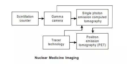
1. Equipment consists of a specialised nuclear imaging camera and a computer. Gamma camera is used which is enclosed in a metallic housing designed to facilitate imaging of specific parts of the body. The camera is mounted on a metal arm that hangs over the examination table.
Operating Principle Of Nuclear Medicine
1. Gamma camera images the gamma (photon) radiation emitted by radioactive compounds. A small dose of radioactive compound is given to patient usually intravenously but sometimes orally so that radioactive material can be localised in specified body organ system. The radioactive compound known as tracer, eventually accumulates in the organ and emits gamma rays. The gamma camera detects the gamma rays, emitted from the body of the patient and works with the computer to produce images which help in measurement and functioning of organs and tissues. Image quality depends on the tracer concentration in the target area and on the emission dynamics of the isotope used. The imaging detector of the camera is made of a sodium iodide crystal where gamma radiation gets absorbed and causes scintillations (tiny flash of light). These are amplified with photo multiplier and the number of scintillations is counted electronically. Spatial localization of the emitting source is achieved with a collimator. The sum of thousands of scintillations creates an image that represents the distribution of radioactivity within an organ or system.
2. The type of tracer to be administered depends on which type of scan is to be performed. Imaging can be done either immediately or after several days. The tracer that is used is determined by what part of the body is under study. It is because some tracers collect in specific organs better than others. Depending on the type of scan, it may take several minutes to several days for the tracer to travel through the body and accumulate in the organ under study.
Single Photon Emission Tomography
1. The computed tomography principles are also being used in nuclear medicine which is then called single photon emission tomography (SPECT). Single photon emission computed tomography is based on a rotating gamma camera. Whereas in CT scan, the image is formed processing x-rays coming out from the body after absorption in the tissues, in SPECT the image is reconstructed using the counted number of emitted photons from the concentration of tracer in the tissues. Like CT scanning, SPECT also uses rotating gamma camera.
Positron-Emission Tomography
1. Positron-emission tomography is a form of nuclear medicine that uses cyclotron during the annihilation of the positron electron pair. Tracers with short life can be produced by cyclotron and computer technology has widely improved. Both have helped in the development of PET. PET measures the difference in travel times of the two quanta’s. This can be used to give the location of tracer where annihilations taking place. PET is an analytical technique that provides a way of making in vivo measurements of anatomical distribution and rates of specific biochemical reactions specially in the brain.
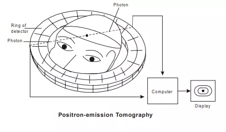
The use of PET to obtain images requires the integration of three components viz (1) radioisotope /tracer (2) PET device and (3) tracer kinetic mathematical models. The positron emitters mostly used are carbon-11, oxygen-15 and nitrogen-13 which have half-life in range of 2 min to 20 min. These tracers are tagged to various metabolically active compounds such as glucose or naturally occurring compounds such as carbon monoxide to image the brain, heart and tumours. The tracers are administered to the patients usually by injection but sometimes by inhalation. Since cyclotron is required to produce positron emitters or tracers, PET has a very high cost.