Mechanics of the Spinal Column
Introduction
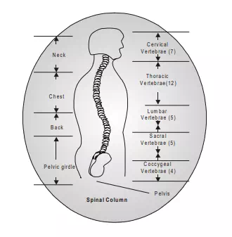
1. The vertical column is also known as spine, spinal column or back bone. It is the control axis of the body. It supports the body weight and transmits it to the ground through the lower limbs. It is the most complex part of the human musculoskeletal system. Its principal functions are to protect the spinal cord, to support the head, neck and upper limbs; to transfer loads from the head and trunk to the pelvis; and to provide flexibility to carry out a variety of movements. It can be divided into five regions viz. cervical (neck), thoracis (chest), lumbar (lower back), sacral and coccygeal regions. The thoracis and lumbar regions of spinal column form the trunk of the body while sacral and coccygeal regions join with pelvis and form parts of pelvic girdle.
Anatomy Of Spinal Column
1. The vertical column is made up of 33 vertebrae’s which include 7 cervical, 12 thoracic, 5 lumbar, 5 sacral and 4 coccygeal. In the thoracic, lumbar and sacral regions, the number of vertebrae corresponds to the number of spinal nerves and each nerve lying below the corresponding vertebrae. In cervical region, then are eight nerves, the upper seven lying above the corresponding vertebrae and the eight below the 7th vertebrae. In the coccygeal region there is only one coccygeal nerve.
2. The vertebrae are also grouped according to their mobility. The moveable or true vertebrae include 7 of cervical, 12 of thoracic and 5 of lumbar vertebrae which have intervertebrae disks in between for facilating rotating movement. Hence these are 24 movable (true) verterbrae and nine unmovable (false) verterbrae which are in sacrum and coccyx region. Twelve thoracic verterbrae have ribs attached to them.
3. There are 24 movable verterbrae and they form amphiarthrodial joints with the fibrocartilaginous interposed between each pair of vertebrae. The fibrocartilaginous discs perform following functions. (1) Sustains loads transmitted from segments above (2) Act as shock absorbers (3) Eliminate bone to bone contact (4) Reduce the effects of impact forces by preventing direct contact between the vertebrae’s. The intervertebrae disc permits articulation of each verterbrae with the adjacent verterbrae in these planes. Hence the entire spinal column functions like a single ball and socket joint. Flexion and extension, lateral flexion and rotation of body is possible due to the structure of spinal column.
4. At the superior end, spinal column has two important joints with head. The atlantooccipital joint is the joint between the first cervical vertebrae (called atlas) and the occipital bone of the head. This is a double condyloid joint (refer chapter 6) The joint permits movement of the head in the sagittal plane and lateral plane. The atlanto axial joint is the joint between the atlas and the axis (first and second verbetrae). This is a pivot joint which permits head to rotate in the transverse plane.
5. The movement of head and neck is provided, controlled and coordinated by a muscle group viz. prevertebral (anterior), hyoids (anterior) sternocleidomastoid (anterior lateral) scalene (lateral), levator scapulae (lateral) suboccipital (posterior) and spleni (posterior). The spine gets its stability from the inter vertebral discs and from the surrounding ligaments and muscles. The discs and ligaments provide passive stability while muscles give active support. The muscles of the spinal column exist in pairs. The anterior portion of spine is connected to abdominal muscles viz. the rectus abdominis, external obligue and internal obligue. These muscles can do flexion & maintain the spine in proper position. There are three layers of posterior trunk muscles viz. the erector spine, the semispinalis and the deep posterior spinal muscle groups. These muscles provide trunk extension as they are located at posterior position of the spine. The effect of gravity is also overcome by these muscles. The quadratus lumborum muscle helps in lateral trunk flexion. The pelvis and lumbar spine is stabilized by this muscle. The lateral flexion of the trunk is carried out by the abdominal and posterior muscles. The rotational movement of the turnk is controlled by the simultaneous action of anterior & posterior muscles.