Compartmental Modelling
Introduction
Compartmental modelling describes the movement of a substance from one compartment to another. Its origins are based on the metabolism of tracer-labelled compound studies in the 1920s. As we will see, compartmental modelling is a special case of physiological modelling, which is covered elsewhere in this book. Primarily, it is concerned with maintaining correct chemical levels in the body and their correct fluid volumes. A compartment can be a volume (or space) or the amount of a substance in a volume. Both representations are commonly used. Here we use the amount of substance in a volume as a compartment, with each compartment assumed to be homogeneous, as described later in the chapter. The process of transfer of substance from one compartment to another is based on diffusion and mass conservation. As shown, compartmental analysis provides a uniform theory that can be systematically applied to many linear and nonlinear systems. While interest in compartmental analysis here is focused on the human body, other engineers and scientists use this technique in studying evolution, carcinogenesis, chemical reactions, infectious disease models, and even semiconductor design and fabrication.
Before investigating compartmental modelling, Fick’s Law of diffusion and osmosis is presented first from basic principles. Next, the volume of a cell and capillary diffusion are discussed. The basics of compartmental modelling are described from simple to more complex models that are increasingly more realistic. Much of the material in this chapter is based on the book by Godfrey.
Solutes, Compartments, And Volumes
When analysing systems of the body characterized by a transfer of a solution from one compartment to another, such as the respiratory and circulatory systems, it is convenient to describe the system as a finite series of interconnected compartments. A solution is defined as a homogeneous mixture of two or more substances in any of the three states of matter: gas, liquid, or solid. Within a solution, we may have a mixture of matter—for instance, solid within a liquid, gas within a liquid, and so on. A solution is described by a component called a solute and another called the solvent. While there are no absolute rules regarding which component is the solute and which is the solvent, we typically call the component in the lesser amount the solute and the other the solvent. For instance, blood is a fluid that consists of 90 percent water with suspended cells such as red blood cells (erythrocytes), white blood cells (leukocytes), platelets, small molecules (i.e., glucose and carbon dioxide), large proteins, and electrolytes (i.e., sodium, calcium, magnesium, potassium). The solute could be a particular protein, and the solvent is the blood minus the protein. The following are some of the readily identifiable compartments in the human body:
· Cell nucleus that is separated from the cytoplasm of the cell
· Internal organelle volumes, such as the mitochondria, endoplasmic reticulum, and so on that are separated from the cytoplasm
· Cell volume that is separated from the extracellular space by the cell membrane
• Interstitium or interstitial volume1 that is separated from the plasma2 by the capillary walls
• Plasma that is separated from the blood
Variables tracked in compartmental analysis are typically quantity or concentration of a solute, temperature, and pressure. Substances of interest are exogenous, such as a drug or tracer, or endogenous, such as glucose or an enzyme or hormone like insulin. Radioactive and stable isotopes3 are used to track the dispersion in a compartmental system and are easily measured. Often a tracer dose of a radioactive isotope is used so the radiation emitted is small and does not interfere with the system. Typically, a tracer dose is less than 1 percent of the total amount of solute in the compartment. High-performance liquid chromatography (HPLC) is used to measure proteins and other macromolecules. Radioimmunoassay is used to measure hormones or proteins, a method based on the immune response of the body to an antigen, which is then bound to an antibody. Other modes of tracking involve injecting a dye (e.g., Evans blue) at one site in the cardiovascular system and measuring the concentration at one or more sites as a function of time.
Fluid in the body is separated into intracellular and extracellular fluid. A typical 70 kg adult contains 42 L of fluid, which is approximately 60 percent of the total body weight. Fluid in the body is tightly regulated so a relatively constant fluid volume is maintained. Intracellular fluid consists of the fluid inside the approximately 75 trillion cells in the body, totaling about 28 L and 40 percent of total body weight. Extracellular fluid consists of two major compartments, the interstitial fluid compartment and the plasma, and two minor compartments, the transcellular fluid and the lymph. The interstitial fluid is 11 L and the plasma is 3 L. The transcellular fluid includes fluids from the synovial, peritoneal, pericardial, intraocular spaces, and the cerebrospinal fluid. These compartments contain approximately 1 to 2 L of fluid. Lymph is the fluid that originates in the interstitial fluid that diffuses into the lymphatic system through lymph capillaries. It returns to the venous plasma after passing through the lymph nodes and has a volume of approximately 1 L. The blood in the circulatory system is a mixture of intracellular and extracellular fluid totalling 5 L of fluid and is 7 percent of total body weight. It consists of the plasma (3 L) and the fluid in the red blood cells (2 L). Table 7.1 summarizes various volumes in the body.
Typically, we work with the plasma compartment rather than the blood compartment, except when dealing with the cardiovascular system. Fluids in the body continually flow from one compartment to another without much change in fluid volume. As will be described more thoroughly in Section 7.3.5, fluid moves from the plasma to the interstitial fluid through the arterial side of the capillary bed and returns from the interstitial fluid to the plasma from the venous side of the capillary bed. Approximately 10 percent of the interstitial fluid does not immediately return to the plasma but moves into the lymphatic system through lymph capillaries by diffusion. The lymphatic system acts like a second parallel circulatory system, with the lymph returning to the plasma after traveling through
TABLE 7.1 Fluid Volumes in a 70 Kg Adult

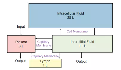
FIGURE 7.1 The compartment volumes of the body. A box depicts the volume. These volumes are tightly controlled by the body through mechanisms that will be described in this chapter. The arrows indicate a flow from one compartment into another (next to the arrows are the types of membranes the fluid must pass through). The rate of flow through a membrane depends on the properties of the membrane. The input includes the fluid ingested. Output is fluid lost from the kidneys, lungs, skin, and sweat, with a small amount lost in the feces. Not shown is the transcellular fluid.
the lymph nodes. The white blood cells in the lymph nodes monitor the lymph and destroy foreign substances to protect the body from disease. Figure 7.1 illustrates the relationships among the compartments.