Although flames and discharges provide a convenient method of excitation, the environment can strongly perturb the sample being studied. Excitation based on broadband-light sources in which the generation of the light is separated from the sample to be investigated provides a less perturbing means of excitation. Higher energy excitation corresponds to shorter wavelengths, but unfortunately, there are not many intense sources of ultraviolet and vacuum-ultraviolet radiation, and so excitation in an electron discharge remains a common method for this portion of the spectrum. (The term vacuum ultraviolet refers to the short-wavelength portion of the electromagnetic spectrum where the photons are energetic enough to excite a typical atom from the ground state to ionization. Under these conditions, the light is strongly absorbed by air and most other substances.)
A typical broadband-light source that can be used for either emission or absorption spectroscopy is a metal filament heated to a high temperature. A typical example is a tungsten lightbulb. Because the atoms in the metal are packed closely together, their individual energy levels merge together; the emitted lines then overlap and form a continuous—i.e., nondiscrete—spectrum. Similar phenomena occur in high-pressure arc lamps, in which broadening of spectral lines occurs owing to high collision rates.
An arc lamp consists of a transparent tube of gases that are excited by an electric discharge. Energetic electrons bombard the atoms, exciting them to either high-energy atomic states or to an ionized state in which the outermost electron is removed from the atom. The radiation that is emitted in this environment is usually a mixture of discrete atomic lines that come from the relaxation of the atoms to lower energy states and continuum radiation resulting from closely spaced lines that have been broadened by collisions with other atoms and the electrons. If the pressure of the gas in the arc lamp is sufficiently high, a large fraction of the light is emitted in the form of continuum radiation.
Light sources that are capable of primarily emitting radiation with discrete, well-defined frequencies are also widely used in spectroscopy. The early sources of spectral emission lines were simply arc lamps or some other form of electrical discharge in a sealed tube of gas in which the pressure is kept low enough so that a significant portion of the radiation is emitted in the form of discrete lines. The Geissler discharge tube, such as the neon lamp commonly used in advertising signs, is an example of such a source. Other examples are hollow cathode lamps and electrodeless lamps driven by microwave radiation. If specific atomic lines are desired, a small amount of the desired element is introduced in the discharge.
Lasers are line sources that emit high-intensity radiation over a very narrow frequency range. The invention of the laser by the American physicists Arthur Schawlow and Charles Townes in 1958, the demonstration of the first practical laser by the American physicist Theodore Maiman in 1960, and the subsequent development of laser spectroscopy techniques by a number of researchers revolutionized a field that had previously seen most of its conceptual developments before the 20th century. Intense, tunable (adjustable-wavelength) light sources now span most of the visible, near-infrared, and near-ultraviolet portions of the spectrum. Lasers have been used for selected wavelength bands in the infrared to submillimetre range, and on the opposite end of the spectrum, for wavelengths as short as the soft X-ray region (that of lower energies).
Typically, light from a tunable laser (examples include dye lasers, semiconductor diode lasers, or free-electron lasers) is directed into the sample to be studied just as the more traditional light sources are used in absorption or emission spectroscopy. For example, in emission (fluorescence) spectroscopy, the amount of light scattered by the sample is measured as the frequency of the laser light is varied. There are advantages to using a laser light source: The light from lasers can be made highly monochromatic (light of essentially one “colour”—i.e., composed of a very narrow range of frequencies). As the light is tuned across the frequency range of interest and the absorption or fluorescence is recorded, extremely narrow spectral features can be measured. Modern tunable lasers can easily resolve spectral features less than 106 hertz wide, while the highest-resolution grating spectrometers have resolutions that are hundreds of times lower. Atomic lines as narrow as 30 hertz out of a transition frequency of 6 × 10¹⁴ hertz have been observed with laser spectroscopy. Because the laser light in a given narrow frequency band is much more intense than virtually all broadband sources of light used in spectroscopy, the amount of fluorescent light emitted by the sample can be greatly increased. Laser spectroscopy is sufficiently sensitive to observe fluorescence from a single atom in the presence of 1020 different atoms.
A potential limitation to the resolution of the spectroscopy of gases is due to the motion of the atoms or molecules relative to the observer. The Doppler shifts that result from the motion of the atoms will broaden any sharp spectral features. A cell containing a gas of atoms will have atoms moving both toward and away from the light source, so that the absorbing frequencies of some of the atoms will be shifted up while others will be shifted down. The spectra of an absorption line in the hydrogen atom as measured by normal fluorescence spectroscopy is shown in Figure 1A. The width of the spectral features is due to the Doppler broadening on the atoms (see Figure 1B.
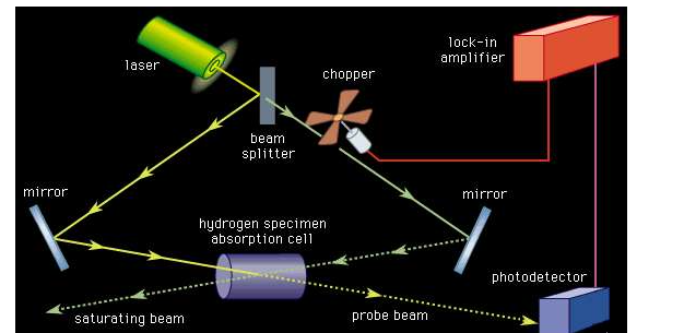
The high intensity of lasers allows the measurement of Doppler-free spectra. One method for making such measurements, invented by Theodore Hänsch of Germany and Christian Borde of France, is known as saturation spectroscopy (see Figure 2). Here, an intense, monochromatic beam of light is directed into the sample gas cell. If the frequency spread of the light is much less than the Doppler-broadened absorption line, only those atoms with a narrow velocity spread will be excited, since the other atoms will be Doppler-shifted out of resonance. Laser light is intense enough that a significant fraction of the atoms resonant with the light will be in the excited state. With this high excitation, the atoms are said to be saturated, and atoms in a saturated state absorb less light.
If a weaker probe laser beam is directed into the sample along the opposite direction, it will interact with those atoms that have the appropriate Doppler shift to be resonant with the light. In general, these two frequencies will be different so that the probe beam will experience an absorption that is unaffected by the stronger saturating beam. If the laser frequency is tuned to be resonant with both beams (this can happen only when the velocity relative to the direction of the two beams is zero), the intense beam saturates the same atoms that would normally absorb the probe beam. When the frequency of the laser is tuned to the frequency of the atoms moving with zero velocity relative to the laser source, the transmission of the probe beam increases. Thus, the absorption resonance of the atoms, without broadening from the Doppler effect, can be observed. Figure 1C shows the same hydrogen spectra taken with saturation spectroscopy.
In addition to saturation spectroscopy, there are a number of other techniques that are capable of obtaining Doppler-free spectra. An important example is two-photon spectroscopy, another form of spectroscopy that was made possible by the high intensities available with lasers. All these techniques rely on the relative Doppler shift of counterpropagating beams to identify the correct resonance frequency and have been used to measure spectra with extremely high accuracy. These techniques, however, cannot eliminate another type of Doppler shift.
This other type of frequency shift is understood as a time dilation effect in the special theory of relativity. A clock moving with respect to an observer appears to run slower than an identical clock at rest with respect to the observer. Since the frequency associated with an atomic transition is a measure of time (an atomic clock), a moving atom will appear to have a slightly lower frequency relative to the frame of reference of the observer. The time dilation can be minimized if the atom’s velocity is reduced substantially. In 1985 American physicist Steven Chu and his colleagues demonstrated that it is possible to cool free atoms in a vapour to a temperature of 2.5 × 10⁻⁴ K, at which the random atomic velocities are about 50,000 times less than at room temperature. At these temperatures the time dilation effect is reduced by a factor of 108, and the Doppler effect broadening is reduced by a factor of 103. Since then, temperatures of 2 × 10⁻⁸ K have been achieved with laser cooling.
Not only have lasers increased the frequency resolution and sensitivity of spectroscopic techniques, they have greatly extended the ability to measure transient phenomena. Pulsed, so-called mode-locked, lasers are capable of generating a continuous train of pulses where each pulse may be as short as 10−14 second. In a typical experiment, a short pulse of light is used to excite or otherwise perturb the system, and another pulse of light, delayed with respect to the first pulse, is used to probe the system’s response. The delayed pulse can be generated by simply diverting a portion of the light pulse with a partially reflecting mirror (called a beam splitter). The two separate pulses can then be directed onto the sample under study where the path taken by the first excitation pulse is slightly shorter than the path taken by the second probe pulse. The relative time delay between the two pulses is controlled by slightly varying the path length difference of the two pulses. The distance corresponding to a 10−14-second delay (the speed of light multiplied by the time difference) is three micrometres (1.2 × 10⁻⁴ inch).