Biological basics of heredity
Basic Cell Structures
By the beginning of the 20th century, it became apparent that a
clear understanding of the processes leading to biological evolution requires
knowledge of how traits are actually transmitted from one generation to
the next. Pursuit of the answer to this question led to the development
of the scientific field known as genetics . Thanks
to Gregor Mendel and other pioneer researchers, we now know that the mechanisms
of genetic inheritance are ultimately responsible for most biological variation
and evolutionary change. In order to comprehend genetics, however, it is
necessary to also understand the biological basis of life. This begins
with the cell .
All plants and animals are composed of microscopic cells, which are the smallest basic organic units capable of carrying out the functions that we normally define as life. These essential cell functions include:
|
1. |
taking in nutrients |
|
2. |
combining the nutrients into substances for cell growth and repair |
|
3. |
reproducing themselves |
|
4. |
excreting waste matter |
Very simple organisms such as bacteria and blue-green algae consist of rudimentary prokaryotic cells. These were the earliest kinds of cells to appear on Earth. More complex single-celled creatures as well as multicellular plants and animals are composed of eukaryotic cells. The description of cell characteristics that follows is for the eukaryotic type cells of which humans are mostly composed.
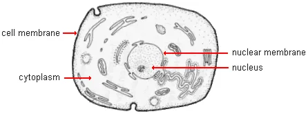
Each of your body's cells is enclosed by a membrane which maintains its integrity. Within is a nucleus with its own surrounding membrane barrier. The nucleus contains most of the genetic material that determines inheritance. Between the nuclear and cell membranes are a number of other specialized cell structures, or organelles. Most of the material outside of the nucleus consists of a water-rich viscous gel referred to as cytoplasm . The cell and nuclear membranes are selectively permeable, which is to say they allow some specific substances to pass through while containing the cell components within. Oxygen, sugar, and other nutrients enter through the cell membrane while useful chemicals produced by the cell as well as carbon dioxide and other waste products of cellular activity leave the cell.
|
|
|
Generalized animal cell |
Red blood cells are an exception to this description in that they do not have a distinct nucleus and nuclear membrane. In this, they are more like the primitive prokaryotic cells of bacteria.
NOTE: There are about 100 trillion cells in each of our bodies. Only 10 trillion of them are human. The other 90 trillion are viruses, bacteria, and other microorganisms. We have evolved symbiotic relationships with most of them. Among other things, they help us digest food and keep us healthy by reducing inflammation and fighting off harmful microorganisms.
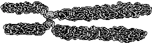
Cells in people and other multicellular organisms reproduce themselves by dividing to form new cells. When the division process begins, long thread-like structures called chromosomes contract and become visible in the nucleus. At other times, chromosomes are, in effect, not visible because of their stretched out thin profile. Chromosomes (literally "colored bodies") were given this name, when they were discovered in 1882, due to the fact that they become visible when stained with a synthetic dye used to enhance the observation of specific cell components.
|
|
|
Contracted human chromosome |
|
|
Species |
|
Number of Chromosomes |
|
|
|
||
|
cat |
38 |
||
|
pig |
38 |
||
|
cow |
60 |
||
|
goat |
60 |
||
|
dog |
78 |
||
|
chicken |
78 |
||
|
|
|||
|
(from "The Promise of
Comparative |
|||
Chromosomes contain almost all of the genetic information that determines inheritance. Different plant and animal species have different shapes and numbers of chromosomes. Humans normally have 46 and our nearest living relatives, the chimpanzees and gorillas, have 48. Having more chromosomes does not necessarily mean that an organism is structurally more complex. For instance, chickens have 78, and there is a fern species that has 1260. More extraordinary still is a microscopic single celled organism named Oxytricha . It has approximately 46 million chromosomes. Having the same number of chromosomes does not mean that two animals are the same species. This can be seen readily from the comparisons in the table on the right.
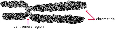
Within a species, different chromosomes are visually distinguishable on the basis of their size and the form of their components. The contracted strands form arms or chromatids . The point of attachment of two or more chromatids is called a centromere .
|
|
|
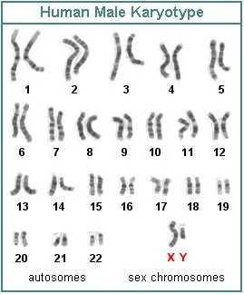
An individual's chromosomes can be photographed when they become visible during the cell division process. Then, all of the pictures of individual chromosomes in a cell may be cut out and organized into a karyotype , such as the one shown on the right. This is a standardized arrangement of the chromosome pictures which allows us to spot major abnormalities that may be characteristic of particular genetically inherited problems, such as Down syndrome . In a karyotype, the chromosomes are placed into homologous pairs. These are pairs that contain genes for the same traits at the same location (or locus) on the chromosomes. However, homologous chromosomes may have different alleles , or alternate forms of the same gene. Homologous chromosomes are paired early in the cell reproduction process.
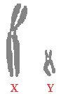
The 46 chromosomes in normal humans consist of 22 pairs of autosomes and one pair of sex chromosomes. The autosomes carry the genes that determine most body characteristics. The sex chromosomes primarily determine sexual traits. If they are X and Y, as in the karyotype shown here, the individual is almost always a male. In contrast, if both sex chromosomes are X's, the gender is female.
In fact, maleness is largely determined by the presence or the absence of a specific gene on the Y chromosome known as the SRY (sex-determining region Y) gene. Beginning in the 5th to 7th week after conception in humans, it triggers other genes that cause the undifferentiated embryonic sex organs to become testes . These, in turn, produce the hormone testosterone which stimulates the development of other masculine physical traits. If the SRY gene is not functioning or absent, an XY individual will very likely develop a female appearing body with breasts and a vagina but will not have a uterus or ovaries and subsequently will be sterile. Instead of ovaries, there will be rudimentary internal testes. Late or partial functioning of the SRY gene can result in individuals being sexually intermediate between male and female. However, this is not the only process that can cause ambiguous sexual characteristics.
Sometimes, in the production of sperm cells, an error can occur that results in a portion of the Y chromosome detaching and becoming part of an X chromosome. If that portion carries the SRY gene and it is inherited, the result can be a chromosomal female (XX) who will develop as a sterile phenotypic male. About 1 in 20,000 normal appearing males are chromosomal females (XX). Roughly the same number of phenotypic females are chromosomal males (XY). Both are sterile.
Sex chromosomes in females and all autosome exist in homologous pairs. That is to say there are 2 copies of each gene (one on each chromosome). These copies may or may not be the same allele for a gene (either may be the dominant or the recessive form). The X and Y chromosomes of males are mostly not homologous--they only share a few genes. Those genes that are found on only the X or the Y chromosome are said to be hemizygous because there is only one copy of each gene. Regardless of whether that gene is a dominant or a recessive allele, it will be expressed in the phenotype of males. You will learn more about this phenomenon in the "Sex Linked Genes" section of this tutorial.
|
|
|
|
Sex Chromosomes |
The X chromosome is much larger than the Y chromosome and has many more genes not related to sexual characteristics. One of the X chromosomes in the cells of human females is partly inactivated early in embryonic life. This is a self-preservation action to prevent a potentially harmful double dose of genes. Recent research points to the "Xist" gene on the X chromosome as being responsible for a sequence of events that silences one of the X chromosomes in females. Apparently, it is the X chromosome from the father that is most likely to be inactivated in developing brain tissues. This may account in part for the "feminizing" of female brains and differences in male and female behavior.
The inactivated X chromosomes become highly compacted structures known as Barr bodies. The presence of Barr bodies has been used at the Olympics and other international sports competitions as a test to determine whether an athlete is a man or a woman. The measurement of testosterone levels has also been used for this purpose.
Every person has about 20,000-25,000 genes in each cell. These genes play a crucial role in determining the specific details of what we are like biologically. The chromosomes are made up of genes. More accurately, the chromosomes are mainly DNA molecules and the genes are sections of them. You will learn more about these molecules later in this tutorial.
The 23 pairs of human chromosomes do not each have the same number of genes. Chromosome 1 is the largest, containing about 8% of all of our genes. Chromosome 21 is the smallest and has only about 1/6 as many genes as chromosome 1.